Sections
|
|
The neck forms a flexible link between the head and the torso, and as such it provides the structural support for the head being both stiff yet flexible, allowing the head to rotate laterally and vertically. It also forms part of the main spinal cord, contains blood vessels leading to the brain and face plus the main respiratory airway (trachea) and digestive intake (oesophagus).
The neck can be divided into 2 main anatomical areas each with their own specific injuries and injury mechanisms.
|
 Figure 13 |
The cervical spine consists of the upper 7 vertebrae, C1 C7, which support the head, and is divided into 2 main vertebrae types.
The Atlas and Axis vertebrae are significantly different from the rest of the vertebrae. The Atlas has no cylindrical load bearing surface and only lateral processes, while the Axis has a much reduced frontal load bearing surface and lateral and posterior processes. As such these bones are significantly weaker than the rest of the spine in compression, shear and bending. To maintain lateral stability between the 2 vertebrae a vertical bone projection in the Axis, the Dens, locates into to a circular opening at the front of the Atlas. It is these bones, which allow the head to rotate, and nod.
The shear strength and bending stiffness of the neck is provided by strong ligaments joining the vertebrae together through the spinal column opening and down the vertebrae spinal processes
In comparison the rest of the cervical spinal C3 C7 is relatively stiff and much stronger.
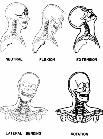
The definitions of motions in the cervical spine is shown below:

|
Dr. A.R. Payne |
S. Patel |
© MIRA 2001 |
|
Project 427519 |
Version 1.1 |